Describe the Types of Oral Lesions
Mouth sores are lesions on any of the soft tissues of the mouth. Varices appear as red blue or deep purple broad-based elevations in oral mucosa.
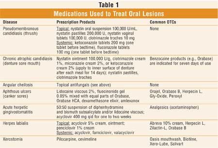
Treatment Of Common Oral Lesions
Lichen planus - benign chronic disease that effects skin and oral mucosa.

. Rubbing from a sharp edge on a broken filling infections systemic conditions associated dermatological diseases and recurrent aphthous ulcers canker sores. A lesion could be a sore wound or any other type of damage tissue caused by injury or disease. Or oral cancer is a type of cancer that originates in the mouth or oral cavity.
Upon compilation of the relevant data oral ulcerative lesions were categorized into three major groups. These lesions may pose a unique diagnostic. You can also ask for the help of the doctor or a specialist.
Oral lesions-reticular or erosive Reticular-interlacing white lines buccal mucosa Erosive-ulcers with erythema and white streaks. 1 Any sores in the mouth that does not heal. An epithelial lined sac that is filled with fluid or semisolid material.
Acute chronic and recurrent ulcers and into five subgroups. This article and photo guide can help. 4 Numbness in or around.
Types of Oral Lesions study guide by kayla_marie_sieburg includes 10 questions covering vocabulary terms and more. The size is usually less than 5 mm. Solitary acute multiple.
Although white lesions constitute. The margins are usually irregular red and painful. There are lesions that extend below the Mucosal surface.
It is solid rough and flat-topped. Diseases of oral soft tissue. 14 rows Large-scale population-based screening studies have identified the.
Ulceration is a commonly presenting sign of a wide spectrum of diseases of the oral cavity involving many etiologic factors. A fluid-filled blister less than 05. Being able to promptly recognize and diagnose oral lesions is critical to heading off several potentially serious conditions.
3 white or rough-textured lesions on the lips or oral cavity. Early examination and prompt diagnosis can aid in prudent. A lesion that is raised like a papule and is greater than 1-2 centimeter 04 to 08 inch in size.
2Describe the types of oral lesions. After you complete the eight-step oral lesion charting then you can start formulating a differential diagnosis. Quizlet flashcards activities and games help you improve your.
Candidiasis - infection caused by yeastlike fungus. Oral lesions can be classified into four groups comprising of ulcerations pigmentations exophytic lesions and red-white lesions. Leukoplakia - white spots on mucosa.
A localized collection of pus in a circumscribed area. This includes the lips cheeks. In this video we talk about how to describe soft tissue and radiographic lesions and the vocabulary used the 8 different approaches to the diagnostic proce.
Acute chronic and recurrent ulcers and into five subgroups. 2 Any lump or swelling on the neck lips or oral cavity. Oral lesions in neonates represent a wide range of diseases often creating apprehension and anxiety among parents.
What causes radiation caries. Upon compilation of the relevant data oral ulcerative lesions were categorized into three major groups. Oral lesions may form individually or multiple lesions.
The most common causes of oral lesions are localised trauma ie. What is the condition frequently seen on the lateral border of the tongue of patients with HIVAIDS. The buccal mucosa is a.
Common Oral Lesions Part I Superficial Mucosal Lesions American Family Physician

Common Oral Lesions Part I Superficial Mucosal Lesions American Family Physician

Pin On Digestive System

Common Oral Lesions Part I Superficial Mucosal Lesions American Family Physician
Comments
Post a Comment